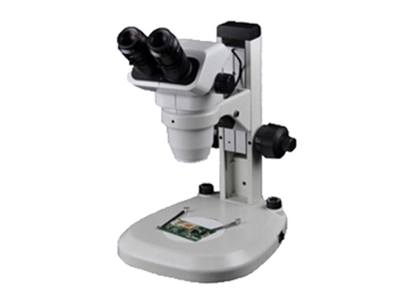
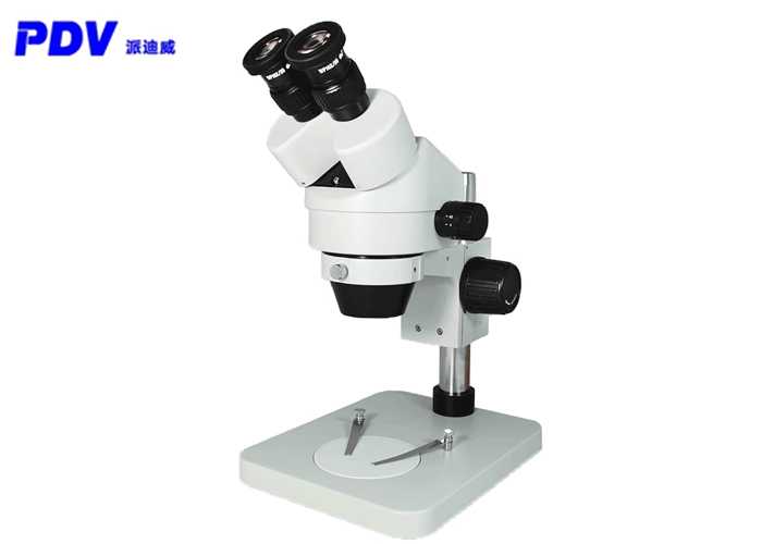
The upright microscopes and inverted microscopes we usually use are collectively called compound microscopes, which can be used to observe smears or slices, cultured living cells, microorganisms in water, or perform microscopic operations.
Bright-field observation
Commonly referred to as bright-field, refers to the illuminating light from the microscope light source through the condenser to illuminate the specimen. After being absorbed by the specimen, the remaining light enters the objective lens and then is seen by the human eye through the eyepiece or captured by the camera through the C interface To. The illumination light path adopts Kohler illumination or critical point illumination.
Blood smears, Gram-stained bacterial smears, stained pathological sections, plant sections, bone grinding films, etc. are all observed in the bright field. These types of specimens are more suitable for observation with an upright microscope. Compared with an inverted microscope, the condenser lens of an upright microscope can be very close to the specimen and has a larger numerical aperture. Therefore, the overall resolution is higher than that of an inverted microscope, and the specimen can be seen more clearly.

The requirements for bright-field observation are mainly clear structure and accurate color. How to ensure clarity and accuracy?
The first is Specimen Preparation. In order to see the clear structure as much as possible, we first pay attention to the question of whether or not to cover the film and how to cover the film in the preparation of the specimen, because different objective lenses have different requirements for whether the specimen is mounted or not.
The "0.17" in the objective lens parameters means that the specimens observed under this objective need to be mounted with a cover glass of thickness 0.17mm; the parameter "0" means that the specimens observed under this objective cannot be mounted with a cover glass, which is suitable for each Kind of smear or polished film; the parameter "-" means that the objective lens has no requirement on whether the objective lens is mounted or not, and both mounted and unsealed specimens can be observed.
For the specimen that needs to be sealed, the specimen shall be immersed in the mounting medium and then covered with a cover glass. It is not allowed to cover the glass directly without mounting medium. The refractive index of the mounting medium is generally close to that of the specimen, and the abrupt change of the refractive index in the imaging optical path is minimized to ensure the best permeability. The refractive index of biological tissue is generally between 1.3 and 1.5, the glass refractive index of the cover glass is about 1.52, and the oil refractive index of the oil lens is 1.518, so the refractive index of the mounted tablet is generally not more than 1.518.
The above information is provided by the motion controller supplier.


