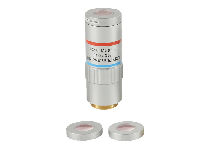
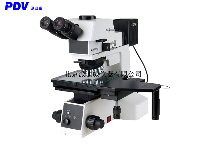
Differential interference microscopy (DIC) is an advanced optical microscopy technique that enables microscopic structures and details in a sample to be observed in a high-contrast manner through the principle of optical interference.
principle
The working principle of the differential interference microscope is based on the interference phenomenon of light. In this type of microscope, a beam of polarized light from a light source is divided into two beams, one affected by structures of different levels or refractive indices in the sample, and the other as a reference beam. The two beams merge again and pass through a polarization beam splitter before entering the objective and eyepiece systems.
When these two beams of light interfere with each other, their phase difference will cause a change in light intensity. This change in light intensity is translated into the image, creating bright and dark areas that increase the contrast of the sample to be viewed. By differential interference, microscopic structures such as cell organs, cell membranes, nuclei, and cytoplasm in samples can be clearly observed without the need for staining or special labeling.
peculiarity
High-contrast observation: Differential interference microscopy provides high-contrast observation, making cells and tiny structures easier to observe in transparent samples.
No staining: Unlike fluorescence microscopy, DIC microscopy does not require staining or labeling of the sample, so the effects of chemical treatment on biological samples can be avoided.
Wide range of application: DIC microscope is suitable for a variety of samples, including biological samples, cells, tissues, liquid crystals, nanomaterials and so on.
Three-dimensional information: The DIC microscope can provide three-dimensional information about the sample because it is able to clearly show the outline and surface of the sample.
Real-time observation: The DIC microscope can be observed in real time and is suitable for observing the dynamic process of biological samples.
Application field
Differential interference microscopy has a wide range of applications in a number of fields, including but not limited to:
Life science: Used to observe biological processes such as cells, cell organs, cell division, and cell movement, and is very important for biomedical research and cell biology.
Materials science: It is used to observe tiny structures, nanoparticles, liquid crystals, etc. in materials, which helps to study the properties and behaviors of materials.
Micropharmacology: Used to study drug interactions with cells to help develop new drugs.
Nanotechnology: For the observation and manipulation of structures and materials at the nanoscale.
Geology: Used to study the microstructure of rocks, minerals and geological samples.


