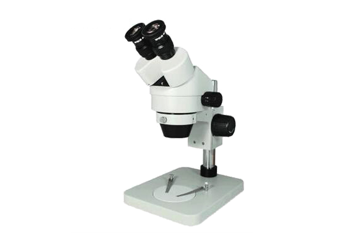
With the advancement of science and technology, people increasingly need to observe the microcosmic world. Microscopes are just such a device. It has broken through the human visual limit and extended it to fine structures that cannot be seen by the naked eye.
Microscopes have been developed since the fifteenth century. From the single-lens microscope designed on the basis of a simple magnifying glass to the complex microscope developed by Zeiss in Germany in 1847, and the appearance of phase contrast, fluorescence, polarized light, and microscopic observation methods, making it more widely used in metals Materials, biology, chemical and other fields.
Microscope objective supplier to share with you: the basic optical principles of microscopes
One. Refraction and refractive index
In a homogeneous isotropic medium, light travels in a straight line between two points. When passing through a transparent object of a medium of different density, a refraction phenomenon occurs, which is caused by the different speed of light propagation in different media. When light that is not perpendicular to the surface of a transparent object enters a transparent object (such as glass) from the air, the light changes direction on its interface and forms a refraction angle with the normal.
two. Lens performance
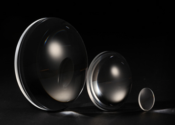
A lens is the most basic optical element that makes up the optical system of a microscope. Objectives, eyepieces, and condensers are composed of single and multiple lenses. According to their different shapes, they can be divided into two categories: convex lenses (positive lenses) and concave lenses (negative lenses).
When a beam of light parallel to the optical axis intersects at a point after passing through the convex lens, this point is called "focus", and the plane passing through the intersection and perpendicular to the optical axis is called "focal plane". There are two focal points. The focal point in object space is called "object side focal point", and the focal plane there is called "object side focal plane". Conversely, the focal point in image space is called "image side focal point". The focal plane is called "image square focal plane".
After passing through the concave lens, the light becomes an upright virtual image, while the convex lens becomes an upright real image. Real images can be displayed on the screen, but virtual images cannot.
3. Principle of Imaging (Geometric Imaging) of Microscope
The reason why a microscope can magnify an object to be inspected is through a lens. Single lens imaging has aberrations, which seriously affects imaging quality. Therefore, the main optical components of the microscope are composed of lenses. From the performance of the lens, it can be seen that only convex lenses can magnify, while concave lenses cannot. Although the objective lens and eyepiece of a microscope are combined by a lens, they are equivalent to a convex lens. In order to understand the magnification principle of the microscope, the five imaging rules of convex lenses are briefly explained:
(1) When the object is outside the focal length of the lens, it will form a reduced inverted real image within the focal length of the image side and out of focus;
(2) When the object is located at the focal length of the lens twice, an inverted real image of the same size is formed at the focal length of the image twice;
(3) When the object is located within the focal length of the lens object and is out of focus, an enlarged inverted real image is formed outside the focal length of the image side;
(4) When the object is located on the focal point of the lens, the image cannot be imaged;
(5) When the object is located within the focal point of the object side of the lens, no image is formed on the image side, and an enlarged upright virtual image is formed on the same side of the lens object side than the object.
The imaging principle of the microscope is to use the laws of (3) and (5) above to enlarge the object. When the object is between F-2F in front of the objective lens (F is the focal length of the object side), a magnified inverted real image is formed beyond the focal length of the objective image side by twice. In the design of the microscope, this image falls within one focal length F1 of the eyepiece, so that the first image (intermediate image) magnified by the microscope objective is magnified again by the eyepiece, and finally in the eyepiece. A magnified upright (relative to the intermediate image) virtual image is formed at the object side (on the same side of the intermediate image) and at the apparent distance (250mm) of the human eye. Therefore, when we are in the microscopy, the image seen through the eyepiece (without additional conversion prism) is the same as the image of the original object in the opposite direction.

Microscope


